نظرة عامة
التصوير بالرنين المغناطيسي للدماغ، ويُسمى أيضًا MRI الدماغ، اختبار تشخيصي يوفر صورًا مفصلة للدماغ. ويمكن أن يلتقط التصوير بالرنين المغناطيسي للدماغ أيضًا صورًا للرقبة والعمود الفقري. وتوضح كل صورة جزءًا أو شريحة منفصلة من الدماغ. وقد يُطلق على التصوير بالرنين المغناطيسي للدماغ كذلك اسم التصوير بالرنين المغناطيسي للرأس أو التصوير بالرنين المغناطيسي للقحف.
أثناء الفحص، تستلقي داخل جهاز طويل على شكل أنبوب يُجري مسحًا لرأسك بينما يلتقط الحاسوب الصور لينشئ صورًا لداخل الدماغ. حيث ينشئ الجهاز صورًا على شكل شرائح لداخل الدماغ - تشبه شرائح الخبز.
يستخدم جهاز الفحص عبر التصوير بالرنين المغناطيسي للدماغ، المعروف أيضًا باسم جهاز MRI الدماغ، جهاز حاسوب وموجات راديوية ومغناطيسًا قويًا جدًا لالتقاط هذه الصور التفصيلية. ولا يسبب هذا الفحص ألمًا. لكنه يُصدِر ضوضاء عالية، وتتطلب بعض الأجهزة الوجود داخل مساحة مغلقة. وقد تكون هذه الأمور غير مريحة لبعض الأشخاص. لا يستخدم التصوير بالرنين المغناطيسي للدماغ الإشعاع كما أنه آمن جدًا لمعظم الأشخاص.
قد تحتاج إلى إجراء تصوير بالرنين المغناطيسي للدماغ لعدة أسباب مختلفة. يمكن أن يُظهر التصوير بالرنين المغناطيسي للدماغ لاختصاصي الرعاية الصحية أي مناطق في الدماغ طرأت عليها تغيرات أو مناطق لا يعمل فيها الدماغ بالشكل الطبيعي. وقد يُستخدم هذا الفحص لتشخيص سبب أعراض مثل الصداع أو التغيّرات في الرؤية أو نوبات الصرع أو فقدان السمع. وتشمل بعض الحالات التي يمكن تشخيصها من خلال التصوير بالرنين المغناطيسي السكتة الدماغية والأورام والتصلب المتعدد.
أجهزة الفحص عبر التصوير بالرنين المغناطيسي للدماغ
جهاز التصوير بالرنين المغناطيسي بقوة 7 تسلا
جهاز التصوير بالرنين المغناطيسي بقوة 7 تسلا
يستخدم جهاز بقوة 7 تسلا المغناطيس الأثقل، ومن ثَم الأقوى. كلما كان المغناطيس أقوى، كانت التفاصيل أكبر في الصور.
توجد عدة أنواع من أجهزة الفحص عبر التصوير بالرنين المغناطيسي، وتُعرف أيضًا بأجهزة MRI:
- التصوير بالرنين المغناطيسي مغلق التجويف، أو ما يُعرف أيضًا بالتصوير بالرنين المغناطيسي التقليدي. يحتوي هذا الجهاز على فتحة في المنتصف، تُسمى التجويف، حيث تستلقي على طاولة بينما يلتقط الجهاز الصور. يلتقط هذا النوع من أجهزة التصوير بالرنين المغناطيسي أوضح الصور وأكثرها تفصيلاً.
- جهاز التصوير بالرنين المغناطيسي واسع التجويف. يتميز جهاز التصوير بالرنين المغناطيسي هذا بفتحة مركزية أوسع من جهاز التصوير بالرنين المغناطيسي التقليدي. ورغم أن المغناطيس لا يزال يحيط بك من جميع الجوانب، إلا أن المساحة الداخلية تكون أكبر. وقد يكون هذا النوع من الأجهزة أكثر راحة للأشخاص ذوي البنية الجسدية الكبيرة أو لمن لديهم رُهاب من الأماكن المغلقة.
- التصوير بالرنين المغناطيسي مفتوح التجويف. لا يُغلق جهاز التصوير بالرنين المغناطيسي هذا عليك بالكامل. حيث توضع المغناطيسات في الأعلى والأسفل، ولكن ليس على الجانبين. ويوفر هذا الجهاز راحة أكبر للأشخاص ذوي البنية الجسدية الكبيرة أو لمن لديهم رُهاب من الأماكن المغلقة.
- التصوير بالرنين المغناطيسي في الوضع العمودي، ويُسمى أيضًا التصوير بالرنين المغناطيسي في وضعية الوقوف أو الجلوس. يتيح لك هذا الجهاز الاستلقاء أو الوقوف أو الجلوس أثناء التقاط الصور. في هذا الفحص، ترتدي ملفًا للرأس يشبه الخوذة. حيث يستقبل الملف الموجات الراديوية وينشئ صورة للدماغ. صُمم التصوير بالرنين المغناطيسي في الوضع العمودي لمساعدة الأشخاص على الشعور براحة أكبر أثناء الفحص. كما أنه يوفر صورًا تُظهر شكل الدماغ في أوضاع مختلفة.
إلى جانب نوع جهاز الفحص، يمكن أيضًا تصنيف أجهزة الفحص عبر التصوير بالرنين المغناطيسي بناءً على قوة المغناطيس. وتسمى وحدة القياس التي تشير إلى قوة المغناطيس تسلا، وتُختصر بالحرف T. وتشمل قوة المغناطيس 1.5 تسلا و 3 تسلا و 7 تسلا. وكلما زاد الرقم، زادت قوة المغناطيس، وزادت التفاصيل في الصور. ويمكن لأي نوع من أجهزة التصوير بالرنين المغناطيسي -سواء كان مغلق التجويف أو واسع التجويف أو مفتوح التجويف أو عمودي التجويف- أن يستخدم مغناطيسًا بأي مستوى من القوة.
في بعض الأحيان، قد يُجرى التصوير بالرنين المغناطيسي للدماغ باستخدام صبغة خاصة تُسمى مادة التباين أو عامل التباين. إذ تُحقن مادة التباين في الوريد قبل الفحص. كما يُستخدم التصوير بالرنين المغناطيسي للدماغ باستخدام مادة التباين إذا احتاج اختصاصي الرعاية الصحية إلى إبراز أنسجة معينة داخل الدماغ. حيث تساعد مادة التباين على تقديم صورة أوضح لبنية الدماغ.
تحتوي عوامل التباين الأكثر شيوعًا المستخدمة في التصوير بالرنين المغناطيسي للدماغ على مادة تسمى الغادولينيوم. وقد يكون بعض الأشخاص لديهم حساسية تجاه الغادولينيوم وقد لا يتمكنون من إجراء التصوير بالرنين المغناطيسي للدماغ باستخدام مادة التباين. كما أن الأشخاص المصابين بأمراض الكلى قد لا يتمكنون من استخدام بعض أنواع عوامل التباين التي تحتوي على الغادولينيوم.
قد تحتاج إلى إجراء تصوير بالرنين المغناطيسي الوظيفي، المعروف أيضًا باسم fMRI. يستخدم التصوير بالرنين المغناطيسي الوظيفي للدماغ الجهاز والطريقة ذاتهما المستخدمين في التصوير بالرنين المغناطيسي التقليدي. ولكن قد يطلب منك الشخص المسؤول عن إجراء التصوير بالرنين المغناطيسي الوظيفي القيام ببعض الأنشطة، مثل تحريك ذراعك أو أداء تمارين ذهنية، كحل مسألة رياضية بسيطة.
يلتقط هذا الفحص النشاط وتدفق الدم في الدماغ أثناء هذه الأنشطة، وليس فقط بنية الدماغ. فعلى سبيل المثال، قد تحتاج إلى عمل تصوير بالرنين المغناطيسي الوظيفي قبل إجراء جراحة في الدماغ وذلك للمساعدة على تحديد المناطق المسؤولة عن الكلام أو الذاكرة في الدماغ. في التصوير بالرنين المغناطيسي الوظيفي، تظهر المناطق التي يزداد فيها تدفق الدم والنشاط في الدماغ أكثر سطوعًا على شاشة الحاسوب.
لماذا يتم ذلك؟
يُجرى التصوير بالرنين المغناطيسي للدماغ للكشف عن عدد من الحالات المرضية المختلفة، ولمعرفة سبب مجموعة متنوعة من الأعراض.
قد يقترح اختصاصي الرعاية الصحية إجراء تصوير بالرنين المغناطيسي للدماغ لمعرفة سبب الأعراض ومنها:
- عدم القدرة على تحريك جزء من الجسم.
- الدوخة.
- الصداع.
- مشكلات في السمع.
- مشكلات في الحديث.
- نوبات الصرع.
- ظهور أعراض بعد إصابة في الرأس.
- مشكلات في الرؤية، مثل فقدان البصر أو الرؤية المزدوجة.
قد يُستخدم التصوير بالرنين المغناطيسي للدماغ لتشخيص حالات صحية مثل:
- النزيف في الدماغ.
- تمدد الأوعية الدموية في الدماغ.
- إصابات الدماغ.
- أورام الدماغ.
- تشكل جلطات دموية في الدماغ.
- الخَرَف.
- الصرع.
- تجمع السوائل في الدماغ، ويُسمى أيضًا الاستسقاء الدماغي.
- مشكلات في الغدة النخامية.
- فقدان الذاكرة.
- الشقيقة والصداع (الصداع النصفي).
- اضطرابات الحركة.
- التصلب المتعدد.
- إصابة الحبل النخاعي.
- السكتة الدماغية.
قد يُستخدم التصوير بالرنين المغناطيسي للدماغ لأسباب أخرى غير تشخيص حالة معينة. تشمل الأسباب الأخرى التي قد تستدعي إجراء التصوير بالرنين المغناطيسي للدماغ ما يأتي:
- رسم خرائط الدماغ. قبل الخضوع لجراحة في الدماغ، قد يحتاج اختصاصي الرعاية الصحية إلى إجراء تصوير بالرنين المغناطيسي للدماغ لتحديد مواقع معينة داخل الدماغ. قد يبحث اختصاصي الرعاية الصحية عن المناطق المسؤولة عن الحركة والكلام والذاكرة حتى يتمكن الجرّاح من توخي الحذر لتجنب هذه المناطق أثناء الجراحة. وقد يُجرى رسم خرائط الدماغ أيضًا قبل العلاج الإشعاعي للدماغ.
- البحث عن تغيّرات في الدماغ. إذا كنتَ قد شُخِصت بالفعل بورم في الدماغ أو أي حالة مرضية أخرى، فقد تحتاج إلى إجراء فحوصات إضافية بالرنين المغناطيسي للدماغ. يمكن أن تحدد هذه الفحوصات ما إذا كان الورم ينمو أم لا. ومع تقدم الأشخاص في العمر، قد يحتاجون إلى إجراء فحوصات التصوير بالرنين المغناطيسي للدماغ للكشف عن وجود انكماش في الدماغ.
- المراقبة. تتطلب بعض العلاجات، مثل العلاجات الجديدة لداء الزهايمر، المراقبة باستخدام فحوصات التصوير بالرنين المغناطيسي للدماغ بهدف تحديد مدى سلامة هذه العلاجات وفعاليتها.
قد يقترح اختصاصي الرعاية الصحية إجراء التصوير بالرنين المغناطيسي للدماغ بدلاً من أنواع أخرى من الاختبارات التصويرية للدماغ، مثل فحوصات التصوير المقطعي المحوسب، لأن التصوير بالرنين المغناطيسي للدماغ يُظهر تفاصيل واختلافات أدق بين أنسجة الدماغ. كما يُفضّل استخدام التصوير بالرنين المغناطيسي للدماغ بدلاً من التصوير المقطعي للأشخاص الذين لا يمكن تعريضهم للإشعاع.
المخاطر
التصوير بالرنين المغناطيسي للدماغ اختبار آمن جدًا بشكل عام ولا ينطوي على مخاطر كبيرة. ولكن قد تزيد بعض العوامل من خطر حدوث مشكلات أثناء التصوير بالرنين المغناطيسي للدماغ.
العناصر المعدنية داخل الجسم أو عليه
نظرًا إلى أن جهاز التصوير بالرنين المغناطيسي يستخدم مغناطيسًا كبيرًا وقويًا، فقد يتطلب الأمر عناية إضافية للأشخاص الذين لديهم معدن على أجسامهم أو داخلها. وتشمل العناصر التي قد تحتوي على معدن:
- أجهزة تنظيم ضربات القلب أو أي من أجزائها.
- مُزيلات الرَّجَفان القلبي القابلة للزرع.
- المسامير أو البراغي.
- الدبابيس الجراحية.
- الدعامات.
- أجهزة التنبيه، مثل المنبهات العميقة للدماغ.
- المعينات السمعية أو الغرسات في الأذن.
- المشابك أو الملفات المستخدمة لعلاج تمدد الأوعية الدموية.
- أجهزة المراقبة المستمرة للغلوكوز.
- الأجهزة المزروعة في الجسم لتوصيل الأدوية، مثل الأنسولين.
- الأجهزة المستخدمة لتوسيع الأنسجة.
- قطع الشظايا أو الرصاص.
- أي جهاز مزروع يحتوي على مغناطيس أو معدن.
- الإكسسوارات التي تُوضع في ثقوب الجسم أو الوشوم أو الكحل الدائم أو مستحضرات التجميل الدائمة الأخرى.
- المفاصل الاصطناعية.
- صمامات القلب الاصطناعية.
قد يجذب المغناطيس الموجود في جهاز التصوير بالرنين المغناطيسي هذه الأجسام المعدنية، ما يؤدي إلى حدوث إصابة. وقد تتطاير الأجسام المعدنية عبر الغرفة نتيجة انجذابها إلى المجال المغناطيسي القوي. كما يمكن أن يُسبب المغناطيس الموجود في جهاز التصوير بالرنين المغناطيسي تسخين الأجسام المعدنية داخل الجسم، وحدوث حروق. لذا، تحدث إلى اختصاصي الرعاية الصحية قبل إجراء التصوير بالرنين المغناطيسي للدماغ لمناقشة أي عناصر قد تؤثر في سلامتك أثناء الإجراء.
إذا كنت مصابًا بالسُمنة أو تخشى الأماكن المغلقة، فقد يصعب للغاية الشعور بالراحة أثناء إجراء التصوير بالرنين المغناطيسي للدماغ. اطلب من فريق الرعاية مناقشة الطرق التي يمكن أن تجعل إجراء التصوير بالرنين المغناطيسي للدماغ أسهل عليك.
مرض الكلى والتصوير بالرنين المغناطيسي للدماغ باستخدام مادة التباين
إذا كنت ستخضع للتصوير بالرنين المغناطيسي للدماغ باستخدام مادة التباين ولديك مرض في الكلى، فسيناقش معك اختصاصي الرعاية الصحية نوع مادة التباين المناسبة لك. عادةً ما تكون أنواع مواد التباين الأحدث التي تحتوي على الغادولينيوم آمنة، لكن قد لا يوصى باستخدام عوامل التباين الأقدم التي تحتوي على الغادولينيوم. قد تكون أكثر عرضة لخطر الإصابة بمرض نادر ولكنه خطير يُسمى التليُّف الجهازي الكلوي المنشأ. ويُسبب هذا المرض زيادة سُمك الأعضاء والجلد والأنسجة الأخرى. وفي حالات نادرة، قد يسبب الوفاة.
إذا كنت تخضع للتصوير بالرنين المغناطيسي للدماغ باستخدام مادة التباين، فمن الممكن أن تُصاب بتفاعل تحسُّسي تجاه مادة التباين. قد يؤدي ذلك إلى الحكة أو ظهور طَفَح. كما يشعر بعض الأشخاص باضطراب في المعدة أو انزعاجات أخرى نتيجة استخدام مادة التباين.
كيف تستعد؟
سيناقش معك اختصاصي الرعاية الصحية ما يجب عليك فعله للاستعداد للتصوير بالرنين المغناطيسي للدماغ. يمكنك عادةً تناول الطعام والشراب دون قيود قبل إجراء التصوير بالرنين المغناطيسي للدماغ. لكن استشر اختصاصي الرعاية الصحية إذا كانت لديك أسئلة حول أطعمة أو مشروبات معينة، مثل المشروبات التي تحتوي على الكافيين أو المشروبات الكحولية. يفترض أن تكون قادرًا على تناول جميع أدويتك اليومية، ما لم يخبرك اختصاصي الرعاية الصحية بعدم تناولها.
يمكنك ارتداء أي ملابس ترغب بها أثناء إجراء التصوير بالرنين المغناطيسي للدماغ، لأنك ستغير ملابسك إلى رداء خاص بالاختبار. يجب أن تهتم بالاستعداد لذلك اليوم وتتجنب ارتداء أو وضع ما يأتي:
- المكياج الذي يحتوي على أي جزيئات معدنية.
- المجوهرات والساعات.
- الدبابيس أو إكسسوارات الشعر المصنوعة من المعدن.
- النظارات الطبية.
- أطقم الأسنان وغيرها من قطع الأسنان المعدنية القابلة للإزالة.
- الإكسسوارات التي تُوضع في ثقوب الجسم.
- المعينات السمعية.
- حمالة الصدر التي تحتوي على سلك معدني.
لا تُسبب عادةً معظم مستحضرات التجميل وحشوات وتقويم الأسنان مشكلات أثناء التصوير بالرنين المغناطيسي للدماغ. ولكن من الممكن أن تؤثر بشكل طفيف في وضوح الصور. تحدث إلى اختصاصي الرعاية الصحية إذا كانت لديك أي أسئلة أو مخاوف.
بشكل عام، لا يُنصح بإجراء التصوير بالرنين المغناطيسي للدماغ للحوامل خلال الأشهر الثلاثة الأولى من الحمل، إلا إذا كان عدم إجراء الاختبار يشكل خطرًا كبيرًا. عادةً ما يكون التصوير بالرنين المغناطيسي للدماغ آمنًا خلال الأشهر الأخيرة من الحمل. وعلى الرغم من عدم وجود إشعاع، فإن الجنين سيكون قريبًا من المجال المغناطيسي القوي للجهاز. يجب على النساء الحوامل عدم الخضوع التصوير بالرنين المغناطيسي للدماغ باستخدام مادة التباين في أي وقت أثناء الحمل. أخبري اختصاصي الرعاية الصحية دائمًا إذا كنتِ حاملاً أو تشتبهين في وجود حمل.
لا تأخذ معك هاتفًا محمولاً أو بطاقات ائتمان أو أي أدوات مغناطيسية أخرى إلى الغرفة لإجراء التصوير بالرنين المغناطيسي للدماغ. فقد تؤثر هذه العناصر في وضوح الصور، وقد يؤدي المغناطيس إلى إتلافها.
نظرًا إلى أن جهاز التصوير بالرنين المغناطيسي قد يصدر أصواتًا عالية جدًا أثناء الاختبار، فستحتاج إلى ارتداء وسيلة لحماية السمع. قد ترغب في إحضار زوج من سدادات الأذن لكي تحافظ على هدوئك وراحتك. وفي بعض المنشآت، قد يعطيك فريق الرعاية سدادات الأذن لارتدائها.
ما يمكن أن تتوقعه
قبل إجراء التصوير بالرنين المغناطيسي للدماغ
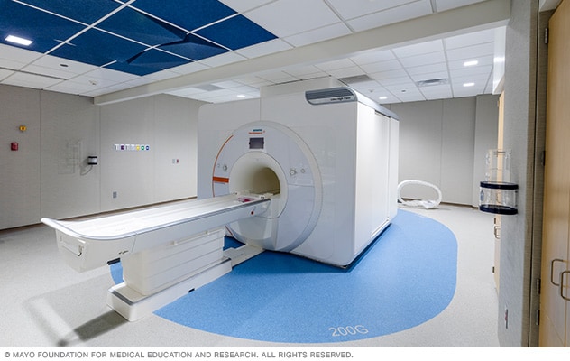
غرفة جهاز الرنين المغناطيسي وغرفة العرض
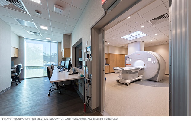
غرفة جهاز الرنين المغناطيسي وغرفة العرض
يوجد جهاز التصوير بالرنين المغناطيسي في غرفة منفصلة عن الفنيِّين الذين يشغلون الجهاز ويلتقطون صور الدماغ. يمكنك التواصل بين الغرفتين من خلال مكبر صوت.
قبل الخضوع للتصوير بالرنين المغناطيسي للدماغ، ناقش مع اختصاصي الرعاية الصحية الأسئلة وأي خطوات يجب عليك اتخاذها للاستعداد. عند وصولك إلى المنشأة، بدل ملابسك إلى رداء المستشفى أو الملابس الطبية الخاصة لإجراء التصوير بالرنين المغناطيسي للدماغ. واتبع تعليمات اختصاصي الرعاية الصحية بشأن الأجسام المعدنية قبل دخول غرفة التصوير بالرنين المغناطيسي للدماغ.
إذا لم تكن متأكدًا أو كانت لديك أسئلة حول وجود أي أجسام معدنية داخل جسمك، فقد تخضع لفحص بالأشعة السينية للتأكد من أن إجراء التصوير بالرنين المغناطيسي للدماغ آمن بالنسبة إليك. وإذا كنت تشعر بالتوتر أو القلق بشأن الفحص أو الوجود في مكان ضيق، فقد يقدم إليك اختصاصي الرعاية الصحية دواءً يساعدك على الاسترخاء. ومع ذلك، يجب أن تكون مستيقظًا أثناء إجراء التصوير بالرنين المغناطيسي.
إذا كان هناك صديق أو أحد أفراد العائلة برفقتك في الغرفة أثناء إجراء التصوير بالرنين المغناطيسي للدماغ، فيجب على هذا الشخص أيضًا اتباع التعليمات المتعلقة بالأجسام المعدنية.
أثناء إجراء التصوير بالرنين المغناطيسي للدماغ
بمجرد دخولك إلى غرفة الفحص، يُطلب منك الاستلقاء على ظهرك فوق طاولة طويلة. ثم ستنزلق الطاولة إلى داخل جهاز التصوير بالرنين المغناطيسي وتخرج منه. يشبه جهاز التصوير بالرنين المغناطيسي التقليدي أسطوانة كبيرة مجوفة أو أنبوبًا مع وجود مساحة في المنتصف للطاولة. ويحتوي الجهاز المحيط بك على المغناطيس.
إذا كنت ستخضع للتصوير بالرنين المغناطيسي المفتوح أو التصوير بالرنين المغناطيسي واسع التجويف أو التصوير بالرنين المغناطيسي في وضع العمودي، فقد يختلف وضعك. اعتمادًا على نوع جهاز التصوير بالرنين المغناطيسي المستخدم، قد لا يحيط بك المغناطيس بالكامل. قد تكون واقفًا أو جالسًا. سيُخبرك فريق الرعاية بكيفية وضع جسمك لإجراء الفحص.
قد تُثَبت ذراعيك وساقيك في مكانهما لإبقائك ثابتًا تمامًا أثناء الاختبار. ثم يضع فريق الرعاية الذي يجري الاختبار ملف تصوير بالرنين المغناطيسي للدماغ حول رأسك. ويشبه هذا الملف الخوذة ويساعد على الحصول على صور أوضح وأكثر تفصيلاً للدماغ.
إذا كنت ستجري تصويرًا بالرنين المغناطيسي للدماغ باستخدام مادة التباين، فسيضع اختصاصي الرعاية الصحية إبرة صغيرة بها أنبوب في وريد اليد أو الذراع. ويحقن اختصاصي الرعاية الصحية مادة التباين عبر الوريد باستخدام هذا الأنبوب. قد تجعلك مادة التباين تشعر بإحساس بالبرودة أو احمرار الجلد في خديك.
تنزلق الطاولة إلى داخل جهاز التصوير بالرنين المغناطيسي، وتكون أنت في وسط المجال المغناطيسي. ويكون اختصاصي الرعاية الصحية الذي يجري الاختبار في غرفة مختلفة. ولكن يوجد نظام اتصال داخلي يتيح لكما السماع والتحدث إلى بعضكما طوال فترة الاختبار.
أثناء استلقائك على الطاولة، قد تسمع أصوات رنين أو دق أو طرقات عالية. وقد يُطلب منك حبس نفَسك في أوقات معينة خلال الاختبار للمساعدة على الحصول على أوضح صورة. قد تشعر أيضًا بإحساس بالدفء على رأسك أثناء الاختبار. إذا كنت تشعر بعدم الارتياح، فأخبر اختصاصي الرعاية الصحية عبر نظام الاتصال الداخلي.
يستغرق التصوير بالرنين المغناطيسي للدماغ عادةً مدة تتراوح بين 30 و 60 دقيقة.
في بعض الأحيان، قد تخضع للتصوير بالرنين المغناطيسي للدماغ أثناء العملية الجراحية. يُستخدم هذا لمساعدة الجراحين على توجيههم أثناء استئصال ورم أو القيام بإجراءات طبية أخرى تتطلب صورًا للدماغ. يُسمى هذا التصوير بالرنين المغناطيسي أثناء العملية، والمعروف أيضًا بالاختصار (iMRI).
كيف يعمل التصوير بالرنين المغناطيسي للدماغ
صور فحص التصوير بالرنين المغناطيسي للدماغ المرجحة بزمن الاسترخاء
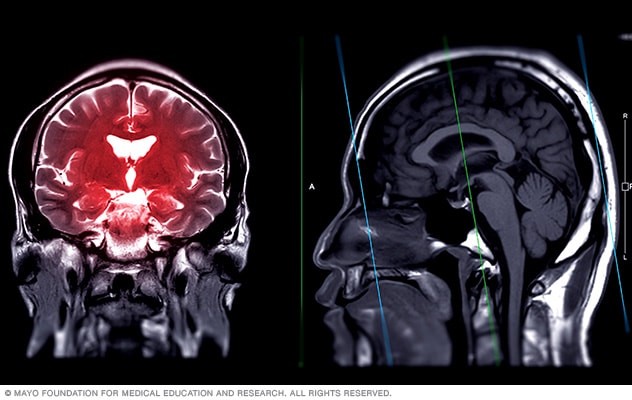
صور فحص التصوير بالرنين المغناطيسي للدماغ المرجحة بزمن الاسترخاء
فحوصات التصوير بالرنين المغناطيسي للدماغ المرجحة بزمن الاسترخاء T2 و T1. يمكن أن تساعد هذه الصور على تشخيص الحالات الدماغية مثل الأورام والتصلب المتعدد والسكتة الدماغية.
يرسل الجهاز موجات الترددات الراديوية إلى داخل الجسم. وتُسمى أنماط موجات الترددات الراديوية والنبضات التي يُنتجها جهاز التصوير بالرنين المغناطيسي باسم التسلسلات. يوفر كل تسلسل نسخة مختلفة قليلاً من طريقة إنتاج الصورة - على سبيل المثال، اختلاف طفيف في التظليل أو الدقة. تشمل بعض أنواع التسلسلات المختلفة المستخدمة في التصوير بالرنين المغناطيسي للدماغ ما يأتي:
- التسلسل المرجح بزمن الاسترخاء T1 مع الغادولينيوم
- التسلسل المرجح بزمن الاسترخاء T1 دون الغادولينيوم
- التسلسل المرجح بزمن الاسترخاء T2
- تصوير الإرواء بالرنين المغناطيسي
- التصوير بالرنين المغناطيسي المرجح بالانتشار
- استعادة الانعكاس المخفف بالسوائل، المعروف أيضًا بالاختصار FLAIR.
"تُثير" موجات الترددات الراديوية ذرات الهيدروجين في الجسم وتدفعها إلى الاصطفاف في خط واحد. وعندما تعود ذرات الهيدروجين إلى أماكنها الطبيعية، فإنها تُطلِق الطاقة. وتختلف كمية الطاقة التي تُطلقها ذرات الهيدروجين حسب نوع النسيج الذي توجد فيه. يقيس الجزء الحاسوبي من جهاز التصوير بالرنين المغناطيسي للدماغ هذه الطاقة ويستخدم المعلومات لتكوين صور للدماغ.
ويرمز الحرف T في هذه التسلسلات إلى زمن الاسترخاء. وزمن الاسترخاء يعني المدة الزمنية التي تستغرقها ذرات الهيدروجين لتصل إلى حالة "الاسترخاء"، وهو ما يعني العودة إلى أماكنها الطبيعية.
بعد إجراء التصوير بالرنين المغناطيسي للدماغ
صورة بالرنين المغناطيسي لورم الدماغ
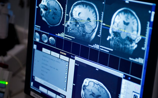
صورة بالرنين المغناطيسي لورم الدماغ
تصوير أورام الدماغ.
بمجرد أن يتأكد أعضاء فريق الرعاية الصحية من حصولهم على الصور التي يحتاجونها للدماغ، يعودون إلى غرفة الفحص لمساعدتك على الخروج من الجهاز. إذا كنت قد خضعت لتصوير بالرنين المغناطيسي للدماغ باستخدام مادة التباين، فإنهم يزيلون الأنبوب الذي وُضع عبر الوريد ويغطون المنطقة بضمادة صغيرة. قد تشعر ببعض الانزعاج أو تلاحظ ظهور بعض الكدمات في موضع الحقن عبر الوريد.
لا تحتاج إلى وقت للتعافي بعد إجراء التصوير بالرنين المغناطيسي للدماغ. ولكن إذا كنت قد تناولت دواءً لمساعدتك على الاسترخاء، فقد تحتاج إلى الانتظار قليلاً حتى يزول تأثيره قبل أن ترتدي ملابسك وتذهب إلى المنزل.
إذا لم تكن قد تناولت دواءً لمساعدتك على الاسترخاء، فيُفترض أن تكون قادرًا على قيادة السيارة بنفسك للعودة إلى المنزل بعد إجراء التصوير بالرنين المغناطيسي للدماغ. ويمكنك العودة إلى ممارسة أنشطتك اليومية المعتادة واتباع نظامك الغذائي مباشرةً بعد إجراء الفحص.
النتائج
بعد الانتهاء من التصوير بالرنين المغناطيسي للدماغ، يقرأ اختصاصي الرعاية الصحية، وغالبًا ما يكون اختصاصي أشعة، النتائج. ويكون اختصاصي الأشعة مُدرَّبًا على قراءة نتائج الاختبارات التصويرية. يستلم اختصاصي الرعاية الصحية الذي طلب إجراء التصوير بالرنين المغناطيسي للدماغ التقرير ويناقشه معك.
ويُخبرك اختصاصي الرعاية الصحية إذا كان كل شيء يبدو سليمًا في الدماغ أو إذا كانت هناك أي مناطق تستدعي القلق. وقد يعني وجود أي نتائج غير طبيعية أنك بحاجة إلى إجراء تصوير إضافي بالرنين المغناطيسي للدماغ أو أي نوع آخر من الاختبارات التصويرية أو اختبارات تشخيصية أخرى. أو قد يرغب اختصاصي الرعاية الصحية في المراقبة والانتظار لمعرفة ما إذا كانت هناك أي تغييرات تظهر مع مرور الوقت.
وإذا تمكن أعضاء فريق الرعاية الصحية من تشخيص سبب الأعراض التي تشعر بها، فسيناقشون معك غالبًا خطة العلاج والخطوات التالية.