Oct. 10, 2023
Core stability and strengthening has become a popular fitness trend. Research has demonstrated that multiple prevalent musculoskeletal injuries, including low back pain, have the potential to disrupt the core and gluteal musculature, or they may be caused by core dysfunction.
In part one of a two-article series, Jane Konidis, M.D., a physiatrist at Mayo Clinic in Rochester, Minnesota, provides an overview of core and gluteal musculature anatomy and muscular slings, and normal and pathological activation patterns. "Having a solid understanding of anatomy and activation patterns in facilitating core stability, especially the role of the gluteus maximus, can help clinicians diagnose and treat the source of many common musculoskeletal injuries," explains Dr. Konidis.
Defining core stability
According to Dr. Konidis, core stability has multiple dimensions, including lumbopelvic stability, neuromuscular control and firing patterns, muscular strength, and gluteal musculature. Functional definitions of core stability also include these elements:
- The ability to control the position and motion of the trunk over the pelvis, allowing optimum production, transfer and control of forces and motion to body segments in integrated kinetic chain activities.
- A stable foundation resulting in proximal stability for distal mobility.
Anatomy components
 عناصر التشريح
تشمل مكونات التشريح الأساسية العضلات واللفافة الصدرية القطنية والروافع التشريحية.
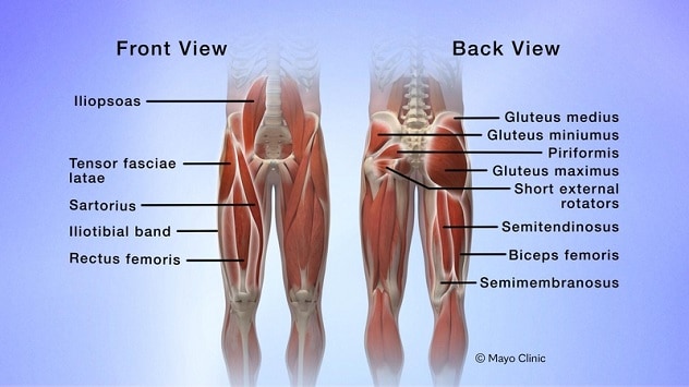
Sustaining core stability involves the integration of multiple components, including muscles, the thoracolumbar fascia and anatomical slings. Dr. Konidis notes that the core is essentially a muscular box that includes local (deep layer) and global (superficial layer) muscles involved in providing lumbopelvic stability. The list of muscles that play a role in core stability includes the abdominal, paraspinal and gluteal muscles; the diaphragm; and the hip girdle and pelvic floor muscles.
Core anatomy components include:
- Muscles. The list of muscles that play a role in core stability includes the abdominal, paraspinal and gluteal muscles; the diaphragm; and the hip girdle and pelvic floor muscles. Local (deep layer) muscles, including the multifidi and transversus abdominis muscles, have origin or insertion points directly or indirectly on the lumbar vertebrae.
- Thoracolumbar fascia. This diamond-shaped web of connective tissue has superficial layer attachments to the gluteus maximus and gluteus medius, the external oblique and latissimus dorsi muscles; and deep-layer attachments to the gluteus medius, erector spinae, internal oblique and serratus posterior inferior muscles.
- Anatomical slings. This network of connected muscles, fascia and ligaments helps produce force vectors and transfer loads through the lumbopelvic region. The four types of anatomical slings are:
- Anterior oblique, including the external and internal oblique, transversus abdominis, linea alba, and contralateral adductor muscles.
- Posterior oblique, including the latissimus dorsi, thoracolumbar fascia and gluteus maximus (contralateral) muscles and the iliotibial band (contralateral).
- Deep longitudinal, including the multifidi and the sacrotuberous ligament.
- Lateral, including the gluteus medius, gluteus minimus and tensor fasciae latae muscle and the iliotibial band.
"Overall, the core musculature helps distribute forces appropriately across the axial skeleton, allowing the generation of maximal strength," explains Dr. Konidis. "Core muscle activation — both local (deep) and global (superficial) — helps create a stable spinal neutral zone and places minimal tension on ligaments.
"These muscles play a critical role in lumbopelvic stabilization," says Dr. Konidis. "Global or superficial layer muscles, including the rectus abdominus and external oblique muscles, span across multiple segments, generating a large amount of torque."
The role of the gluteus maximus
Dr. Konidis notes that the gluteus maximus plays an often overlooked but significant role in facilitating core stability and hip extension and in stabilizing the pelvis. This is especially true during trunk rotation or during a shift of center of gravity. This large muscle can affect the stability of the lower back, the femoral head and possibly even knee extension via its attachments into the iliotibial (IT) band.
Research originating among athletic trainers and the bodybuilding community has gradually influenced medical professionals' interest in developing a better understanding of the role of the gluteus maximus in providing core stability and preventing injuries. Studies suggest that weaknesses and delayed firing of the gluteus maximus are often present in individuals with lower back pain or lower extremity instability.
"In a healthy-functioning individual, the gluteus maximus has the potential to be the most powerful and largest muscle in the body," explains Dr. Konidis. "But several factors can cause dysfunction in this muscle, leading to reduced or delayed activation. Factors causing this dysfunction can include prolonged sitting, hip flexor muscle group overactivity, and pain from local or distal injuries. Many people have 'flat butt' syndrome from sitting for so long."
Muscle activation patterns
During movement, a normal healthy sequence in activation of core musculature occurs. Injury or pain, both locally and distally, can perturb these sequences, which can in turn lead to excessive strain on synergists and further injuries. And when muscle inhibition occurs, it can lead to the perception of pain via feedback loops, changes in motor programming and disturbed patterns of activation.
"Any weak links in this chain can put excess strain on muscles and lead to injury in both the lower and upper extremities," says Dr. Konidis. "Injuries related to biomechanical overload can occur through synergistic dominance, when a muscle with a similar function takes on a load for an injured muscle.
"If a prime mover such as the gluteus maximus is weak or inhibited — with lower activation strength — synergistic muscles must then compensate," explains Dr. Konidis. "If a muscle is tight or shortened, that muscle may be recruited earlier, which may throw off the normal pattern of activation. This means that muscle inhibition patterns and muscle weakness or tightness can alter normal movement patterns. We also know that multiple factors can contribute to muscle inhibition. Any time that loads are increased and altered at the other segments and passive structures, these changes can be a recipe for pain generation and further injury," says Dr. Konidis.
In part two of this article series, Dr. Konidis outlines a practical approach to evaluating and treating problems related to gluteal muscle dysfunction.
For more information
Mayo Clinic Physical Medicine and Rehabilitation Medical Professionals Resources
Refer a patient to Mayo Clinic.