Feb. 09, 2022
Mayo Clinic uses ultrasound-guided aspiration to determine the presence of prosthetic joint infection (PJI) after total hip arthroplasty. Orthopedic surgeons apply a technique developed at Mayo Clinic that yields sufficient synovial fluid for culture in most patients, including individuals with high body mass index (BMI).
Aspirations of hip arthroplasties have traditionally been performed in fluoroscopy suites. However, fluoroscopy doesn't show soft tissues and fluids as clearly as does ultrasound. The ultrasound-guided procedure also avoids radiation exposure and provides greater convenience for patients.
"The ultrasound procedure can be easily combined with a patient's visit to the orthopedic clinic because both appointments are in the same location," says Suzanne M. Tanner, M.D., an orthopedic surgeon at Mayo Clinic in Rochester, Minnesota.
PJI is a potentially devastating complication of total hip arthroplasty, leading to complex revision surgery, prolonged antibiotic therapy, morbidity and the risk of death. Determining the presence of PJI is challenging, as there is no single, accurate test.
At Mayo Clinic, all patients undergoing revision of total hip arthroplasty typically have ultrasound-guided aspiration to check for PJI. "Serum inflammatory markers aren't totally reliable. Around 4% of people with normal inflammatory markers still have an infection," Dr. Tanner says. "Short of operating to obtain tissue specimens, obtaining fluid specimens for culture is the most important test to determine if a joint is infected."
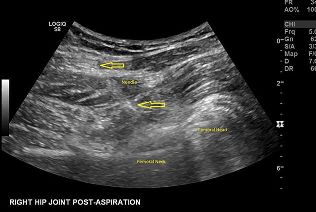
Ultrasound image of hip aspiration

Ultrasound image of hip aspiration
Ultrasound image of hip aspiration shows needle placement (yellow arrows) in relation to labeled parts of a hip prosthetic.
The major challenge of ultrasound-guided aspiration is obtaining enough synovial fluid for accurate analysis. Mayo Clinic uses a stepwise approach. Initially, the aspiration needle is placed at the neck of the prosthesis. If sufficient fluid isn't obtained, the needle is directed laterally and just past the femoral component neck. Lavage also might be used to draw out fluid.
Another challenge is possibly impaired visualization in patients with high BMIs. Although a high proportion of individuals undergoing ultrasound-guided hip aspirations at Mayo Clinic have BMIs above 30, sufficient fluid is usually obtained, including in patients with BMIs above 40.
As a result, ultrasound-guided aspiration at Mayo Clinic generally provides highly accurate diagnosis of infection. "With a meticulous technique and exacting protocols, we are able to obtain useful fluid with a low contamination rate," says Holly J. Duck, M.D., an orthopedic surgeon at Mayo Clinic's campus in Minnesota. "That success helps our surgical colleagues to determine the best treatment for our patients."
"It's somewhat rare for orthopedic surgeons rather than radiologists to test for PJI," Dr. Tanner says. "At Mayo Clinic, we have a sufficiently large space and patient volume to provide this type of care."
For more information
Refer a patient to Mayo Clinic.