April 20, 2018
Complex structural deformities of the foot and ankle, especially those caused by neurological disorders or trauma, present some of the most time-intensive and technically challenging cases for orthopedic surgeons and involve some of the most difficult recoveries for patients. Many of these deformities result from a combination of muscular, anatomical and neurological dysfunction.
For example, in the hereditary sensorimotor peripheral neuropathy Charcot-Marie-Tooth (CMT) disease, some muscles become weaker than others. This leads to an imbalance of forces across the foot and ankle, which in turn leads to deformity. Over time, the deformity progresses, causing pain, difficulty wearing shoes, and sometimes skin breakdown, nonhealing ulcers and infections.
The goal in treating deformity in the foot and ankle, including CMT, is to prevent complications and achieve a plantigrade, stable foot that allows reasonable function. This can be accomplished surgically by realigning the foot using osteotomies or fusions and balancing the forces across the foot by way of soft tissue releases or tendon transfers.
A common approach for a symptomatic, relatively moderate deformity in CMT is first to osteotomize the calcaneus and first metatarsal to bring about a more plantigrade foot. In a more severe deformity, the shape of the foot is corrected through a hindfoot fusion or triple arthrodesis. The foot is then balanced using soft tissue releases and tendon transfers, such as an Achilles tendon lengthening, plantar fascia release, peroneus longus to peroneus brevis tendon transfer and transfer of the tibialis posterior tendon to the dorsal foot. In this way, the foot is made plantigrade with osteotomies or fusions, and it is balanced through the soft tissues.
"Processes such as Charcot-Marie-Tooth have classic presentations — a cavus, high-arched foot, clawed toes and ankle laxity," says Daniel B. Ryssman, M.D., an orthopedic surgeon at Mayo Clinic's campus in Rochester, Minnesota. "But others are unique. It's important to examine patients thoroughly to see what is working and what isn't. Surgeons must rely on X-rays, a careful physical exam and sometimes nerve conduction studies to determine what is causing the deformity. Then they can start planning what to do."
Dr. Ryssman says treatment is dictated by the location and type of deformity, disease severity, and the presence of infection and other comorbidities as well as by patient goals and degree of social support.
"Some patients simply aren't good candidates for surgery because of comorbidities, or the deformity may just be too great to fix. I can usually straighten the foot, but if the patient still can't use it because of skin loss or a recurring infection, surgery isn't in his or her best interest. In such cases, the only real options are to leave the foot alone or perform an amputation," he explains. "But other considerations are critical, too. What is the patient's chief complaint, and what is the social situation at home? Is there support? You have to really listen to the patient before deciding what to do."
In straightforward cases, the deformity can be corrected in a single stage. Patients remain in a cast for 10 weeks and then transition to a boot. A month or so later, they begin wearing shoes, with full recovery expected within six months to a year.
Other deformities are not suitable for acute correction and internal fixation, have a significantly increased risk of complications or poor soft tissue coverage, or are multiplanar or rigid. These may require a staged surgery involving percutaneous osteotomy, significant releases and external fixation with a device such as the Taylor spatial frame.
The frame, which consists of two or more rings connected to six telescopic struts that can be lengthened or shortened, allows gradual postoperative correction of the deformity by changing strut lengths according to a computer-generated treatment plan.
"By making 1-millimeter adjustments to the struts every day, the rings are slowly repositioned and the deformity is gradually corrected at a pace that allows the skin and soft tissue to heal," Dr. Ryssman says. "This can take up to six months, and although it is challenging for patients, the technique has helped correct very severe deformities."
A case study
Patient's feet at initial visit

Patient's feet at initial visit
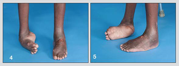
Images from the initial visit of patient with severe bilateral equinocavovarus foot deformity
One such case involved a Somali man in his early 20s, who first saw Dr. Ryssman in April 2012. A gunshot to the young man's spine had caused neurological damage leading to severe bilateral equinocavovarus foot deformity.
"This young patient was ambulating on the tops of his feet and would have to swing his legs through his crutches," Dr. Ryssman says. "The deformity was fixed and rigid, and I initially thought there was no way to treat him, but then decided we might be able to work with the frame.
"I was concerned, however, because he didn't have family here, although an aunt was willing to help. Eventually, after three or four meetings, I decided to perform the surgery on one foot, which turned out to be extremely complex. The normal boney architecture was so twisted it was difficult to apply the fixator."
Adjusting to the fixator was a challenge for the patient, too, who fainted the first time he saw it, Dr. Ryssman says. He gradually grew used to it, however, and by the time he left the hospital was proficient at making the strut adjustments.
Patient's left foot corrected

Patient's left foot corrected
First foot corrected
Final correction of patient's feet

Final correction of patient's feet
In images of the final correction, both of the patient's feet are plantigrade.
A year later, Dr. Ryssman surgically corrected the other foot, again using the external fixator. Today, both feet are plantigrade, and the young man is able to walk in braces without pain.
"His feet are not normal, but there is a significant difference, and we achieved our goal," Dr. Ryssman says. "Proceeding with the correction was a very difficult decision. The patient was essentially living alone and his English was imperfect. I wondered whether I was doing the right thing for him given his social situation and whether this might be too much for him. But it ended up making a tremendous difference in his life.
"These cases are all multifactorial; they require a great deal of planning and a long recovery for patients, but at the one-year mark, when they are recovered and walking straight, it's very satisfying and sometimes quite emotional to see patients and their families so happy."