July 21, 2023
Increasing interest in the clinical use of ultrahigh field 7-Tesla (7-T) MRI focuses largely on managing epilepsy and multiple sclerosis (MS). Mayo Clinic neuroradiologists have expanded clinical use of 7-T MRI beyond these typical applications — routinely using the advanced technology to guide care for patients with neurodegenerative disorders, cerebrovascular disease, amyotrophic lateral sclerosis (ALS) and other neurological conditions.
"7-T imaging is technically very challenging, which has limited its adoption in clinical medicine. But we've devoted a lot of resources, expertise and time to tackling these challenges," says Erik H. Middlebrooks, M.D., a neuroradiologist at Mayo Clinic in Jacksonville, Florida. "As a result, we're developing new tools to assess patients with unprecedented accuracy."
Mayo Clinic's campus in Florida performs 7-T MRI for about 1,200 patients a year — the highest clinical use of 7-T MRI in the United States. Mayo Clinic neuroradiologists have developed several 7-T MRI sequences that facilitate the technology's wider use.
"7-T MRI can be a game changer. It's hard to overstate the impact it has on our patients' lives."
Focal cortical dysplasia
3D edge-enhancing gradient-echo (EDGE) — an MRI sequence developed by Mayo Clinic — can improve the detection of focal cortical dysplasia. 7-T MRI can potentially boost that improvement, but only if challenges involving image-contrast uniformity are overcome.
Mayo Clinic has developed a new imaging strategy that does just that. As described in the March 2023 issue of the American Journal of Neuroradiology, the protocol combines 7-T EDGE and a variation of magnetization prepared rapid gradient echo (MPRAGE) called MP2RAGE.
Localización de la lesión
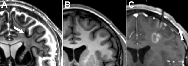
Localización de la lesión
A. Una resonancia magnética frontal de 7 teslas con realce de bordes revela una displasia cortical focal (flecha) responsable de un largo historial de convulsiones del paciente. B. En una resonancia magnética de 3 teslas, no se observó la lesión de manera concluyente. C. Se extirpó la lesión mediante una terapia térmica intersticial inducida por láser. El paciente no ha vuelto a tener convulsiones.
The new strategy is yielding positive results. Dr. Middlebrooks cites a 20-year-old man with a long history of medication-refractory focal seizures that occurred about three times daily. Although inpatient monitoring had revealed focal onset, no lesions were apparent on multiple prior 3-T MRI scans.
"MRI at 7-T using the new sequence revealed a dysplasia with excellent correlation to the localization recorded on inpatient monitoring," Dr. Middlebrooks says. The patient had laser interstitial therapy to ablate the lesion.
"This young man had dealt with seizures since childhood, with no answers. The condition affected every aspect of his life. He was unable to drive and had increasing difficulty with school and other issues. Over a year after surgery, he has not had a single seizure and will soon be getting his master's degree."
Neurovascular conditions
3-T MRI generally finds no evidence of intracortical infarctions or microhemorrhages after transient ischemic attack (TIA).
Mayo Clinic has demonstrated that 7-T MRI can localize those abnormalities. The discovery, to be published in The Neuroradiology Journal, is the first report of intracortical localization of microhemorrhages in patients with TIA.
"Detecting microinfarctions on 7-T imaging can allow more confidence in attributing a patient's symptoms to a vascular etiology. 7-T imaging also has potential to prognosticate risks of future infarctions or hemorrhages."
Studies have also indicated the potential of 7-T MRI to provide clearer images in time-of-flight (TOF) magnetic resonance angiography (MRA). But clinical use of 7-T MRA has been limited by factors such as high specific absorption rate and increased acquisition time.
Detalles muy precisos
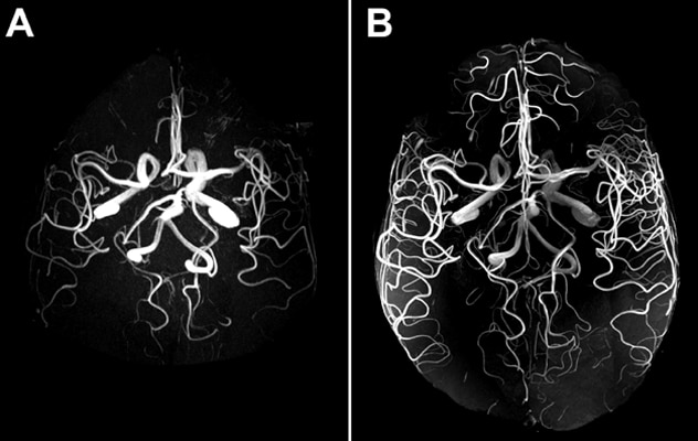
Detalles muy precisos
A. Angiografía por resonancia magnética de tiempo de vuelo sin contraste de 3 teslas de las arterias intracraneales. B. Una angiografía por resonancia magnética de tiempo de vuelo de 7 teslas permite visualizar los vasos más distales y los más pequeños.
Mayo Clinic has demonstrated successful clinical application of 7-T TOF MRA using compressed sensing. "7-T MRA can provide images at 300 microns or smaller, which provide exquisite details that are pertinent to diagnosis," Dr. Middlebrooks says.
The application is described in a study published in the June 2023 issue of The Neuroradiology Journal. In a separate report, published in the March 2023 issue of Radiology, Mayo Clinic neuroradiologists describe their use of 7-T MRI to characterize a 4-millimeter aneurysm in a patient's left ophthalmic artery.
Cognitive disorders
7-T MRI is paving the way for new approaches to transient global amnesia (TGA) and leukoencephalopathy.
In a case report to be published in The Neuroradiology Journal, Mayo Clinic researchers describe how 7-T MRI revealed a residual lesion in a patient's hippocampus eight months after she experienced TGA. The imaging also showed evidence of gliosis and volume loss.
"This finding challenges the traditional mantra of TGA as a fully reversible condition with no long-term imaging findings. TGA may have more lasting effects than previously believed."
7-T MRI can also reveal signs of CSF1R-related leukoencephalopathy. In a case study published in the December 2022 issue of Neurology, 7-T MRI revealed characteristic frontal lobe calcifications in a 24-year-old woman who had screening for a family history of the condition. The patient experienced no symptoms, and routine neurological examination and 3-T MRI showed no abnormalities.
"The improved sensitivity of 7-T MRI benefitted treatment decision-making," Dr. Middlebrooks says. "We were able to refer the patient to a clinical trial."
Other applications
Mayo Clinic has also developed 7-T MRI protocols for assessing:
- Trochlear nerve pathology. Described in the February 2023 issue of American Journal of Neuroradiology, the protocol was used to review 100 nerves in 50 individuals undergoing 7-T MRI for epilepsy care. At least two segments of the trochlear nerve from the brainstem to the cavernous sinus were identified in 100% of cases.
- Parkinson's disease. This protocol uses multi-echo gradient echo sequences with magnetization transfer pulse at 7-T to image neuromelanin in the substantia nigra. Diminished neuromelanin in that region is a marker of Parkinson's disease. As explained in the November 2022 issue of Radiology, the sequence can also reveal elevated iron deposition, another histopathological feature of Parkinson's disease.
- ALS. Using ultrahigh resolution 7-T MRI of the motor cortex, Mayo Clinic neuroradiologists can detect the characteristic histologic changes that occur within the middle cortical layers related to degeneration of the cell bodies of corticospinal and callosal neurons. This promising biomarker not only improves early diagnosis but also may allow patients earlier access to emerging therapies.
Future directions
Captar pequeñas venas y lesiones
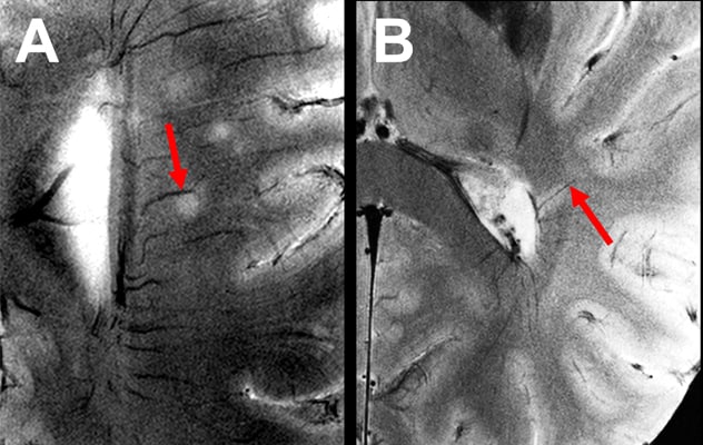
Captar pequeñas venas y lesiones
La resonancia magnética de 7 teslas permite detectar con precisión pequeñas venas asociadas a lesiones de la sustancia blanca. A. Se determinó que un paciente remitido por esclerosis múltiple tenía migraña con aura. La resonancia magnética de 7 teslas demostró una localización excéntrica de la vena relacionada con la lesión (flecha), típica de las lesiones de la sustancia blanca por enfermedad vascular de vasos pequeños. B. Un segundo paciente no cumplía los criterios tradicionales de diagnóstico de la esclerosis múltiple. Sin embargo, en una resonancia magnética de 7 teslas, se observó una vena central patognomónica dentro de una lesión (flecha), lo cual permite diagnosticar la esclerosis múltiple.
Mayo Clinic is conducting the largest study to date of 7-T MRI for assessing individuals with MS.
"7-T MRI is one of the best diagnostic tools we have for MS," Dr. Middlebrooks says. "It has led to major changes in how some patients are managed. We've also had patients referred to us with a diagnosis of MS and found that the cause of their symptoms was entirely different."
Mayo Clinic neuroradiologists and physicists work closely with the 7-T MRI system's manufacturer to optimize use of the technology. "We are aggressively pursuing extended applications of 7-T MRI," Dr. Middlebrooks says. "It takes a lot of multidisciplinary effort, resources and expertise. But we are committed to helping this technology benefit more patients."
For more information
Tao S, et al. Edge-enhancing gradient-echo MP2RAGE for clinical epilepsy imaging at 7T. American Journal of Neuroradiology. 2023;44:268.
Lakhani DA, et al. 7T MRI in transient ischemic attacks: Have we only seen the tip of the iceberg? The Neuroradiology Journal. In press.
Lakhani DA, et al. Clinical application of ultra-high resolution compressed sensing time-of-flight MR angiography at 7T to detect small vessel pathology. The Neuroradiology Journal. 2023;36:235.
Desai A, et al. Cerebral aneurysm imaging at 7T with use of compressed sensing. Radiology. 2023;306:e221277.
Singh RB, et al. Chronic hippocampal subfield damage in transient global amnesia revealed by 7T MRI: All is not reversible? The Neuroradiology Journal. In press.
Dulski J, et al. Novel application of 7T MRI in CSF1R-related leukoencephalopathy. Neurology. 2022;90:1110.
Kumar T, et al. High-resolution 7T MR imaging of the trochlear nerve. American Journal of Neuroradiology. 2023;44:186.
Lakhani DA, et al. 7-T Neuromelanin and R2* MRI in Parkinson Disease. Radiology. 2022;305:296.
Refer a patient to Mayo Clinic.