June 03, 2023
Neuroscientists have long understood that cells in the precentral gyrus send signals directly to the peripheral nervous system to generate voluntary movement. It's also known that these cells are principally organized as a topologic map of the body. This correspondence of an area on the cortex to a specific region of the body is called somatotopy.
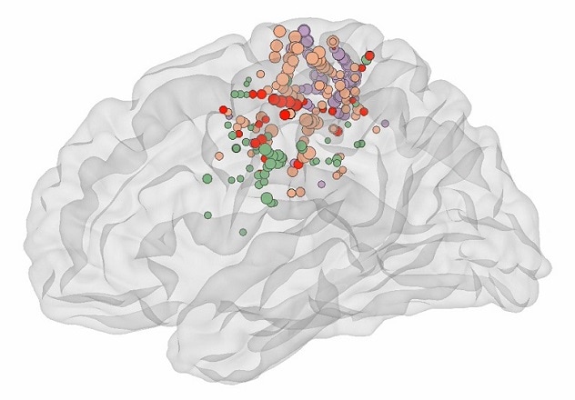
Mayo Clinic scientists have discovered that this somatotopic map extends into the depths of the central sulcus, an area lacking electrophysiological evidence for control of voluntary movements. What's more, the somatotopy is interrupted by an area deep in the central sulcus that the scientists call the Rolandic Motor Association area (RMA).
The team set out simply to characterize the primary motor cortex electrophysiologically throughout its 3D volume using penetrating stereoelectroencephalography (SEEG) depth electrodes. The electrodes are implanted in individuals with drug-resistant epilepsy in order to localize the areas of seizure onset. The technology allows for simultaneous measurements from superficial and deep brain areas.
The patients performed a simple block-designed task of randomly interleaved tongue, hand or foot movements — contralateral to SEEG array — with rest in between. Electromyography recorded muscle activity from each body area. Simple analysis of broadband power changes in SEEG — a correlate of neural cell population activity — extended the classic somatotopic representation of individual body parts into the sulcal depths of the precentral gyrus.
The researchers expected to find only the classic body-based map along the anterior bank of the central sulcus.
The Rolandic Motor Association area (RMA) in the central sulcus

The Rolandic Motor Association area (RMA) in the central sulcus
Dots show areas of the brain controlling foot (purple), hand (orange) and tongue (green) movements — as well as a fourth area (red) active in all three movement types, which the researchers call the RMA.
"Instead, we found a region interrupting the otherwise somatotopic representation in the depths of the central sulcus at its midlateral aspect, active during all three movement types," says Michael A. Jensen, an M.D./Ph.D. candidate at Mayo Clinic in Rochester, Minnesota. "Because the RMA isn't plainly related to any single movement function, we believe that it's likely an association area that helps to coordinate complex movements."
The scientists note that the discovery of the RMA area is highly disruptive to the standard understanding of the precentral gyrus as a purely somatotopic brain region. Mayo Clinic's work has strong potential to change how neuroprostheses are implanted and to alter stimulation paradigms and planning for surgical resections.
"This basic finding of an association area deep in the central sulcus forces us to rethink our understanding of how the circuitry of movement is organized," says Kai J. Miller, M.D., Ph.D., a neurosurgeon at Mayo Clinic's campus in Minnesota. "We hope emerging science about this RMA area's role will allow improved therapies for our patients."
For more information
Jensen MA, et al. A motor association area in the depths of the central sulcus. Nature Neuroscience. In press.
Refer a patient to Mayo Clinic.