Overview
A SPECT scan is a type of imaging test that uses a radioactive substance and a special camera to create 3D pictures. This test is also known as single-photon emission computerized tomography.
While many imaging tests show what the internal organs look like, a SPECT scan can show how well the organs are working. For instance, a SPECT scan can show how well blood is flowing to the heart; what areas of the brain are more active or less active; or what parts of the bone are affected by cancer.
Products & Services
Why it's done
Some of the most common uses of SPECT are to help diagnose or monitor brain disorders, heart problems and bone disorders.
Brain disorders
A SPECT test creates a detailed, 3D map of the blood flow activity in the brain, which can help see which parts of the brain are being affected by:
- Clogged blood vessels. SPECT scanning can find issues with blood flow in the brain. It can help diagnose or check on vascular brain disorders, such as moyamoya disease, a condition in which the arteries in the brain become blocked or narrowed.
- Seizure disorders. A SPECT scan can help diagnose and treat seizure disorders, such as epilepsy. It does this by pinpointing the area of seizure activity in the brain.
- Parkinson's disease. In rare cases, a healthcare professional may suggest a SPECT scan called a dopamine transporter scan (DaTscan) to help confirm a diagnosis of Parkinson's disease. Parkinson's disease is a progressive neurological condition that affects movement.
Some medical institutions may use SPECT scanning to help check other brain conditions, such as dementia or head trauma.
Heart problems
Because the radioactive tracer highlights areas of blood flow, SPECT can check for:
- Clogged coronary arteries. If the arteries that feed the heart muscle become narrowed or clogged, the parts of the heart muscle served by these arteries can become damaged or even die.
- Weak pumping action. SPECT can show how completely your heart chambers empty during contractions.
Bone disorders
Areas of bone healing usually light up on SPECT scans, so this type of test is being used more often to help diagnose hidden bone fractures. SPECT scans also can diagnose and track cancer that has spread to the bones. It also can help find sites for bone biopsy.
More Information
Risks
For most people, SPECT scans are safe. If you have an injection or infusion of radioactive tracer, you may experience:
- Bleeding, pain or swelling where the needle was inserted in your arm.
- Very rarely, an allergic reaction to the radioactive tracer.
Be sure to tell your healthcare team or radiation technologist if there's a possibility you're pregnant or if you're breastfeeding.
Risks of radiation
Your healthcare team uses a small amount of radiation to perform a SPECT scan, and the test is not associated with any long-term health risks. Talk to someone on your team if you're concerned about your exposure to radiation during a SPECT scan.
How you prepare
How you prepare for a SPECT scan depends on your situation. Ask your healthcare team whether you need to make any special preparations before your SPECT scan.
In general, you should:
- Leave metallic jewelry at home.
- Tell the technologist if you're pregnant or breastfeeding.
- Bring a list of all the medicines and supplements you take.
What you can expect
During the test
SPECT scan

SPECT scan
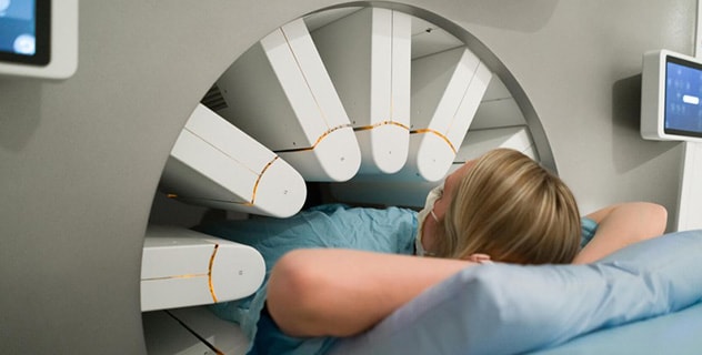
During a SPECT scan, your healthcare team positions you on a table. Then the SPECT machine rotates around you, taking pictures of internal organs and other structures highlighted by the radioactive tracer in your body.
SPECT scans involve two steps: receiving a radioactive injection, called a tracer, and using a SPECT machine to scan a certain area of your body.
Receiving a radioactive substance
You'll receive a radioactive substance through an intravenous (IV) infusion into a vein in your arm. The tracer dose is very small, and you may feel a cold sensation as it enters your body. You may be asked to lie quietly in a room for 20 minutes or more before your scan while your body absorbs the radioactive tracer. In some cases, you may need to wait several hours or, rarely, several days between the injection and your SPECT scan.
Your body's more active tissues will absorb more of the radioactive substance. For instance, during a seizure, the area of your brain causing the seizure may hold on to more of the radioactive tracer. This can pinpoint the area of the brain causing your seizures.
Undergoing the SPECT scan
The SPECT machine is a large circular device containing a camera. It can detect the radioactive tracer absorbed by your body. During your scan, you lie on a table while the SPECT machine rotates around you. The SPECT machine takes pictures of your internal organs and other structures. The pictures are sent to a computer that uses the information to create 3D images of your body.
How long your scan takes depends on the reason for your procedure.
After the test
Most of the radioactive tracer leaves your body through your urine within a few hours after your SPECT scan. You may be told to drink more fluids, such as juice or water, after your SPECT scan. This helps flush the tracer from your body. Your body breaks down the remaining tracer over the next few days.
Results
SPECT scan results

SPECT scan results
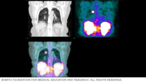
SPECT scan results can be in color or shades of gray. The varying shades or colors show which cells in the body are absorbing more or less of the radioactive tracer. This scan includes images of the kidneys, liver and spleen.
A radiologist or healthcare specialist with advanced training in nuclear medicine will study the results of your SPECT scan and send them to your healthcare team. Pictures from your scan may show colors that tell your team what areas of your body absorbed more of the radioactive tracer and which areas absorbed less. For instance, a brain SPECT image might show a lighter color where brain cells are less active and darker colors where brains cells are more active. Some SPECT images show shades of gray, rather than colors.
Ask your healthcare team how long to expect to wait for your results.
Clinical trials
Explore Mayo Clinic studies of tests and procedures to help prevent, detect, treat or manage conditions.
Jan. 05, 2024