Jan. 31, 2015
A major advance in neurosurgery has been the development of new endoscopic techniques for skull base tumors. Skull base tumors pose significant challenges due to their close proximity to important neurovascular structures and potential extension into neural canals or the cavernous sinus. Surgical resection can be associated with significant morbidity, including damage to the optic nerve and leakage of cerebrospinal fluid.
Jamie J. Van Gompel, M.D., of the Department of Neurologic Surgery at Mayo Clinic in Rochester, Minnesota, says, "Everything we do centers around maximal safe resection of tumors, limiting comorbidity to patients, and trying to get them back to the normal work or family life they previously had." Mayo's experience with skull base lesions ranges from rare types — esthesioneuroblastomas, chordomas and chondrosarcomas — to the more common meningiomas, pituitary tumors, craniopharyngiomas and sinonasal malignancies.
Dana Erickson, M.D., of the Division of Endocrinology, Diabetes, Metabolism, and Nutrition at Mayo Clinic in Rochester, Minnesota, says: "All of our patients with pituitary tumors who go to surgery undergo either the endoscopic-microscope approach or the expanded endoscopic approach."
Endoscopic endonasal approach
Endoscopic endonasal approach
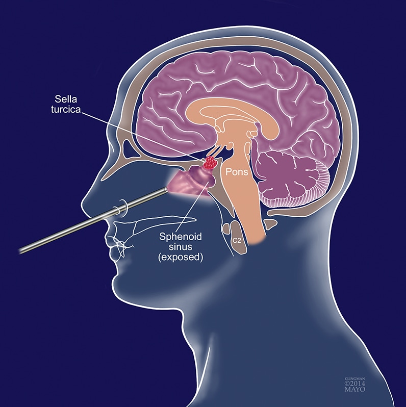
An endoscopic endonasal approach to the resection of sellar and parasellar tumors allows visualization of areas in the sella that could not be seen previously and enhances the opportunity for a complete pituitary tumor resection.
Dr. Van Gompel adds: "The advantage of the expanded endoscopic approach is that we can now visualize areas in the sella that we could not see previously, enhancing the opportunity for a complete pituitary tumor resection. Unlike the microscope, which focuses light narrowly on the tumor — it is like looking through a keyhole in a door — the endoscope works more like a flashlight, bringing the light and visualization to the tumor.
"The surgery is usually performed by both a neurosurgeon and an otorhinolaryngologist. There are two expert sets of eyes, and often intraoperative discussion about what's best for the patient. In addition, endoscopes offer different angles of vision, and once the bulk of a pituitary tumor is resected, the endoscope can be advanced into the sella and we can look around the corner."
This technique is of particular value in patients with large tumors with significant suprasellar or cavernous extension.
Dr. Erickson comments: "For our patients with hormone-secreting tumors, like corticotroph tumors causing Cushing's syndrome or somatotroph tumors causing acromegaly, complete resection at the first operation is key to successful long-term cure."
Dr. Van Gompel adds: "The expanded use of the endoscope has been made possible by technological advances in neurosurgical techniques, improvements in light intensity and high-definition imaging."
Conclusion
Dr. Erickson concludes: "The range of experience and expertise at Mayo Clinic allows patients to receive care appropriate to their individual needs. The development of endoscopic techniques for the management of sellar and parasellar tumors has been a major advance in our pituitary practice."
For more information
Van Gompel JJ. Skull Base Surgery. Medical Professional Video Center. Mayo Clinic. 2014.